Here is my favourite essay on the structure of plant cell, explained with the help of suitable diagrams.
Protoplasm:
Protoplasm is the essence of life Huxley described it as ‘the physical basis of life’. All the characteristics of living beings like metabolism irritability, growth and reproduction are the external manifestations of the internal changes of the protoplasm. The protoplasmic mass inside a single cell is called the protoplast. So it is the unit of protoplasm and is the metabolically active component of the cell.
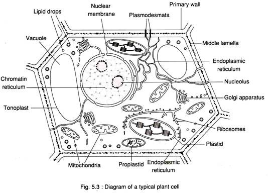
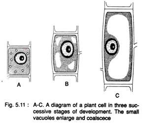
It consists of three parts – the cytoplasm, the nucleus and the vacuole. The ground substance which resides immediately beneath the cell wall and surrounds the vacuole is the cytoplasm. The young cell contains very small vacuoles and, as the cell enlarges, the vacuoles coalesce to form a large one. With the increase in cell size the protoplasmic content does not increase in proportion to the vacuole size and, as a result, a thin cytoplasmic layer surrounds the vacuole.
This thin layer is known as the primordial utricle. The vacuole is bound by a membrane known as the tonoplast separating the vacuolar sap or cell sap from the cytoplasm. The cytoplasm in its periphery is, again, bound by a membrane known as the cell membrane or plasma membrane.
The soluble portion of the cytoplasm in which the cell inclusions remain embedded is known as the cytosol. The extensive membranous system which traverses the entire cytoplasm is called the endoplasmic reticulum (ER). It consists of a ramified network of interconnected, membrane-bound tubules and vesicles.
Essential organelles occurring in all eukaryotic cells are the mitochondria, which are more numerous and somewhat smaller and serve as the centres of intracellular oxidation. They are generally spherical to ellipsoidal, and are commonly about 1 µm thick and 1-3 µm long. Like plastids they are bound by a pair of membranes and are also semi-autonomous in function.
Plastids are the second largest semi- autonomous cell inclusions enclosed by a double membrane. They are confined to the plant kingdom. They may be coloured or colourless. They are typically lens-shaped with a diameter of up to 10 µm. There are about 200-400 such plastids per plant cell.
The nucleus is the largest particulate inclusion, which is spherical with a diameter in the range 6-8 µm. A perforated double membrane surrounds the nucleus. This membrane is continuous with the ER membranes. The nuclear membrane surrounds a network of chromatin matter known as chromatin reticulum which remains embedded in a nuclear sap known as the karyolymph.
Also, there is a dark spherical body attached to the reticulum known as the nucleolus which is responsible for ribosome formation. The nucleus is an autonomous structure. It controls all the cell functions. It is also responsible for the transmission of hereditary characteristics from generation to generation.
The Golgi apparatus is a component of the endomembrane system of the cell and has the property of being both particulate and membranous. The endomembrane system of the cell consists of cisternae which are stacks of flattened vesicles, Golgi bodies or dictyosomes closely associated with ER membrane and surrounded by tiny spherical vesicles, and Golgi apparatus The inter-associated dictyosomes which function synchronously, form a Golgi apparatus.
The most numerous of all the subcellular particles are the ribosomes, occurring in the cytoplasm, either free or attached to ER membranes and nuclear membranes. They are also found in chloroplasts and mitochondria. The ribosomes in the cytoplasm are slightly larger than those in the chloroplasts and mitochondria. They are more or less spherical in shape and made up of approximately equal amounts of RNA and protein and are actively involved in protein synthesis.
Microbodies consitute another group of particulate inclusions. ‘Microbody’ is a morphological term for a distinct class of organelle. It includes peroxisomes and glyoxysomes, the former occurring in photosynthetic cells and the latter restricted to cells of the endosperm or cotyledons of fat-storing seeds. The microbodies are roughly spherical and single-membrane structures with a diameter of 0.2 -1.5 µm.
Another debatable particulate inclusions are the spherosomes which are restricted to the cells of lipid-storing tissue. They are also single-membrane spherical structures with a diameter of 0.4- 3 µm.
All the living cells in higher plants are interconnected by thin strands of cytoplasm known as plasmodesmata. Typically they are cylindrical holes through the cell wall. Each hole is lined by a cylinder of plasmalemma, the lumen of which is filled with cytoplasm and it often contains a minute tubule — known as the desmotubule.
There are spaces around each living cell called intercellular spaces. Initially, in the meristematic region, the cells remain in contact with each other in such a way that there are no intercellular spaces. But during differentiation and development the primary walls separate at the corners, forming intercellular spaces.
In mature tissues these spaces become interlinked forming an intercellular space system which ends at the stomatal openings. So, through the stomatal openings, the whole system gets air circulation by diffusion, ensuring an adequate supply of O2 and CO2 to the tissues for their metabolic activities.
The existence of plasmodesmata and intercellular spaces subdivides the plant body into two major compartments, known as the symplast and apoplast, respectively. The former is the living part of the plant made up of the interconnected protoplasts bound by single continuous plasmalemma.
The latter is the non-living part of the plant external to the plasmalemma and composed of cell walls, the intercellular spaces and the dead cell lumens such as the xylem vessels. Both the compartments are utilised for the transport of materials through the plant.
Cell Membrane:
The protoplast of plant cells is separated from their environment by the plasma membrane. The structure and function of the plasma membrane in plants is fundamentally similar to that in animals, fungi, and bacteria, but specific knowledge of many aspects of the plant plasma membrane is lacking. The composition of the lipid components and the properties of the proteins vary from membrane to membrane, on which membrane’s unique functional characteristics depend.
The plant cell membrane has a number of important functions, such as to mediate the transport of solutes into and out of the protoplast, to coordinate the synthesis and assembly of cell wall microfibrils, and to translate hormonal and environmental signals involved in the control of growth and differentiation.
The first important hypothesis of the structure of biological membranes was proposed by H. Davson and J. Danielli in 1935. This hypothesis says that membranes contain a continuous hydrocarbon phase contributed by the lipid components of the membrane. The hypothesis was later modified by J. D. Robertson into the Unit- Membrane Hypothesis.
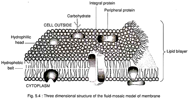
The unit membrane was proposed to consist of a bilayer of mixed polar lipids, with their hydrophobic tails oriented inward to form a continuous hydrocarbon phase and their hydrophilic heads oriented outward.
Each surface was thought to be coated with a monomolecular layer of protein molecules with the polypeptide chains in extended form. It was suggested that the unit membrane is about 8.0 to 9.0 nm thick and that the lipid bilayer is about 6.0 to 7.0 nm thick.
The most satisfactory model of membrane structure at present is the dynamic fluid-mosaic model put forward by Singer and Nicholson in 1972. This model postulates that amphipathic lipids (e.g., phospholipids, glycolipids, sterols) are arranged in a bi-layer to form a fluid, liquid- crystalline matrix or core, in which individual molecules can move laterally.
This movement gives the bi-layer fluidity, flexibility and characteristically high electrical resistance and relative impermeability to highly polar molecules. Globular proteins form a mosaic on the matrix. They may be partially embedded on either side or may completely penetrate the membrane. This mosaic is not fixed or static, because the proteins are free to diffuse laterally in two dimensions.
The fluid-mosaic model accounts satisfactorily for many features and properties of biological membranes:
(i) It explains the widely different protein content per unit area,
(ii) It explains the variation in the thickness of different types of membranes,
(iii) It explains the electrical properties and permeability of membranes, and
(iv) It also explains the movement of some proteins in the cell membrane.
Functions:
The plasma, membrane has at least three major functions:
(i) It is highly specialised for the transport of a variety of substance into and out of the cell,
(ii) It has an important role in the synthesis and assembly of cell wall microfibrils,
(iii) The plasma membrane is charged with the reception of a variety of external signals and the translation of these signals into various biochemical and physiological responses through de-repression of a number of genes.
Plastids:
Plastids are organelles characteristic of plant cells. They vary in shape, size, and pigmentation pattern. There are three main types of plastids – chloroplastids, chromoplastids, and leucoplastids. Chloroplastids are green due to the presence of chlorophyll as a main pigment.
Chromoplastids are usually yellow, orange, or red — because of the presence of carotenoid pigments. They occur in petals, in some ripe fruits, and in some roots (e.g., carrot). Leucoplasts are colourless non-pigmented plastids, usually located in tissues not exposed to sunlight. Usually they store food such as starch (amyloplastid), proteins (proteinoplasts), and fats (elaioplasts).
Starch, phytoferritin (an iron- protein complex) and lipids together form globules called plastoglobuli, which may be present in all types of plastids. Leucoplasts, on exposure to light, are converted to chloroplasts (e.g., in the potato tuber).
The proteinoplasts of Helleborus corsicus were observed to initiate as amyloplasts; later the starch dis-appears and the main product of the plastid becomes protein. All types of plastids are derived from minute bodies called pro- plastids. Plastids and pro-plastids in young cells multiply by division.
The chloroplast usually has a characteristic lens shape with a length of 4-10 µm.
There are three major structural parts of the chloroplast:
(i) An envelope consisting of two membranes with an enclosed space,
(ii) the mobile stroma containing the soluble enzymes for CO2 fixation, protein synthesis, and starch storage, and
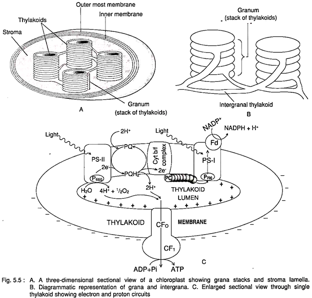
(iii) the highly organised internal lamellar membranes stacked together to form a grana which performs the function of light energy capture. Each stack is a disc or sac-like vesicle and is termed thylakoid (in Greek, sac-like) by Wilhelm Menke (1960) (Fig. 5.5).
The outer envelope is 50-80 Å thick. It is differentially permeable and regulates the passage of sugars and other carbon intermediates, NADPH/NADP+, ATP and ADP in and out of the chloroplast. Synthesised starch is retained inside as storehouse of carbon skeleton for further use.
The stroma is a proteinaceous matrix containing about 50% ribulose 1, 5-bisphosphate carboxylase/oxygenase (Rubisco). There are small droplets rich in lipids, particularly plastoquinone close to the thylakoid membranes. These droplets are called plastoglobuli. The starch grains remain close to the thylakoids and are usually ovoid and up to 1.5 µm in length.
The stroma also contains ribosomes (70S) attached to the outer surface of the thylakoid membranes. It also contains 20-50 identical histone-free circular DNA molecules with a circumference of 36-62 µm. A few chloroplast proteins are coded by chloroplast DNA The large subunit of Rubisco is known to be coded by a chloroplast gene while the small subunit is coded by a nuclear gene.
The higher plant chloroplasts contain two types of thylakoid, the large or stroma thylakoid and the small or grana thylakoid. There may be 40-60 grana per chloroplast and usually 5-20 thylakoids per granum. Each thylakoid encloses an internal space called thylakoid lumen (Fig. 5.5).
The membranes between the grana are perforated. The thylakoid system constitutes a single complex cavity separated from stroma by the thylakoid membrane system. Built into the thylakoid membranes are pigment systems and electron carriers which carry out the light reaction of photosynthesis.
Mitochondria:
Mitochondria are called the power houses of cell as they serve as sites of ATP synthesis.
Generally, a plant cell contains several hundred simple mitochondria. They are spherical, ellipsoidal or rod-like structures. Mitochondria are specifically stained by the dye Janus green B. The mitochondria move freely in the streaming cytoplasm and appear to divide and coalesce; they undergo considerable changes in shape, being globular, cylindrical or branched.
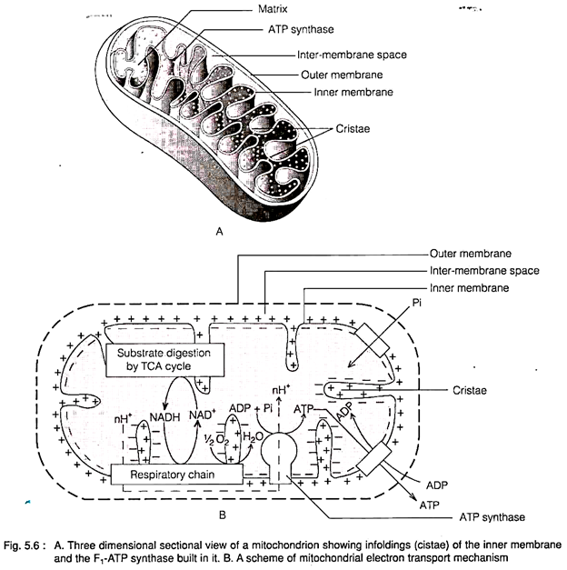
Mitochondria have a double-membrane envelope. There is an aqueous matrix of solutes, soluble enzymes, and the mitochondrial glucose encircled by the inner membrane which is larger than the outer one and remains infolded to form sac-like cristae of variable shape and number, usually with a narrow neck (Fig 5.6).
There is an inter-membrane space in- between the two membranes. The mitochondrial matrix is finely granular and highly proteinaceous. The matrix contains a fibrillar region with DNA known as nucleoid. It also contains ribosomes and calcium containing granules.
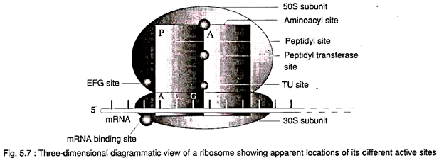
The ribosomes have a sedimentation coefficient of 70S. The ribosomes are composed of a large subunit (50S) and a small subunit (30S) Higher plant mitochondrial DNA is a circular molecule of 30µm length with a molecular weight of 70 x 106.
Ribosomes:
The term ribosome was first introduced by Roberts in 1958. They are ribonucleoprotein particles and are essential machinery for protein biosynthesis. Green plant cells have 80S ribosome in the cytoplasm and 70S (sedimentation coefficient) ribosomes in the mitochondria and chloroplastids.
The diameter of 80S ribosome is 20-22 nm, and the particle weight is about 4.0 megadaltons. The ribosomes are composed of two subunits, one of which is larger than the other (Fig. 5.7). The larger subunit is 60S and the smaller one is 40S. Plant ribosomes contain 25S and 16S rRNA.
The ribosomal RNAs are highly folded, and double helical stems alternate with single- stranded loops. The small subunit fits over the ends of the large subunit in such a way as to form a tunnel between the particles.
This tunnel could accommodate the m-RNA, tRNA and protein synthesis factors. The shape of the ribosomes mainly depends on the concentration of Mg2+ ions in the medium. Removal of Mg2+ causes dissociation and unfolding of the subunits.
Endoplasmic Reticulum:
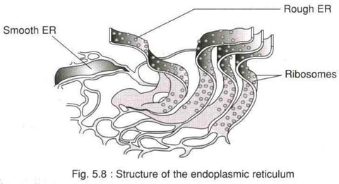
Electron microscopic studies reveal that plant cells, especially secretory cells, contain an extensive membrane network of interconnecting tubules and cisternae (flattened sacs) traversing the entire cytoplasm. This network is termed the endoplasmic reticulum (ER) and was first described in plant cells by Buvat and Carasso (1957) (Fig. 5.8).
The ER membrane is somewhat thinner than the plasma membrane measuring 5.6 nm in thickness. In the metabolically active cell the endoplasmic reticulum consists of interconnected parallel cisternae associated with ribosomes towards the cytoplasmic face of the membranes. This is known as the rough endoplasmic reticulum (RER) – a major site of protein synthesis.
If the ER membranes do not bear ribosomes they are called smooth-ER (SER). SER serves as a major site of lipid synthesis and membrane assembly. RER, on the other hand, serves as the working bench for the synthesis of proteins. The ER membranes are rich in phosphatidyl choline and phosphatidyl ethanolamine and lesser amounts of phosphatidyl inositol and phosphatidyl glycerol.
The endoplasmic reticulum has got the following important functions:
i. It functions as a communication or transport system throughout the cell, mediating the intracellular transport of small and large molecules.
ii. The ER tubules often end at plasmodesmata and thus play a role in intercellular communication.
iii. It plays an important role in cellular metabolism.
iv. It is the principal site of membrane synthesis and thus contributes to the formation of other organelles.
Golgi Apparatus:
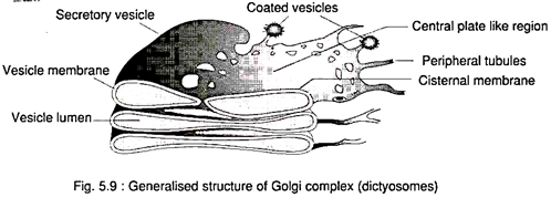
The Golgi apparatus was discovered in 1898 by the Italian scientist Camillo Golgi. It consists of a system of stacks of flat, smooth-walled circular cisternae or saccules made up of unit membrane (Fig. 5.9). Each stack is termed Golgi body or dictyosome. Each dictyosome contains 5-8 cisternae.
The margins of the cisternae may be vesicular, fenestrated or tubular in shape. The vesicles are budded-off from the cisternal margins. In some cases rod-shaped elements in parallel arrangement have been seen in the very narrow spaces between the individual cisternae Golgi apparatus also contains coated or smooth surfaced vesicles of various types.
It is a pola structure. The proximal pole (forming face) of each dictyosome remains associated with the nuclear envelope of ER in a characteristic manner. The opposite pole (maturing face) however becomes more like the plasma membrane. The inter-associated dictyosomes together form the Golgi apparatus. The number of dictyosomes per Golgi apparatus ranges from 0-25,000.
The Golgi apparatus is a component of the endomembrane system of the cell and appears as intermediate between ER and cell membrane.
Golgi apparatus performs the following main functions:
1. The Golgi bodies are concerned with secretion processes. It functions in the packaging of materials for export to the cell’s exterior. They are mainly involved in the secretion of sugars (in nectar secretion), polysacchandes (cell-wall material, mucilage), and polysaccharide protein complexes (certain mucilages).
2. They are also responsible for the formation of a new plasma membrane to support growth or to replace the lost one.
Cytoskeleton:
Throughout the cytosol there is a three- dimensional network of filamentous proteins called the cytoskeleton. It gives the scaffold structure for the positioning and movement of the organelles. In plant cells the cytoskeleton consists of microtubules, microfilaments, and intermediate filaments of fixed diameter and variable lengths.
The microtubules are hollow filaments of 25 nm diameter, microfilaments are solid, having 7 nm diameter, and the intermediate filaments are helically coiled fibres of 1 nm diameter. Microtubules and microfilaments are made up of globular proteins. The protein polymers of tubulin are the components of microtubules.
Microfilaments are composed of globular actins or G-actins each of which is composed of a single polypeptide. Intermediate filaments are composed of linear polypeptide monomers of various types like lamins, keratins etc.
The cytoskeleton is involved in a variety of processes like intracellular movements such as cytoplasmic streaming, mitotic movements secretory vesicle transport, cell plate formation, and cellulose micro-fibril deposition etc.
Nucleus:
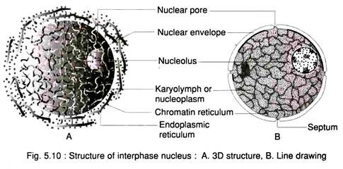
The nucleus is usually more or less spherical, though nuclei with other shapes have also been observed. The nucleus is surrounded by a nuclear envelope and contains the nuclear matrix called nucleoplasm and one or more nucleoli (Fig. 5.10).
The nucleoplasm contains chromatin reticulum consisting of deoxyribonucleic acid (DNA) and proteins. The DNA and protein complex in the chromosomes having affinity to basic dyes is called chromatin. In the interphase the chromosomes are uncoiled and can not be distinguished and form a network called the Chromatin reticulum.
At this phase some condensed and deeply stained chromatin masses are often seen in the reticulum. This chromatin is called heterochromatin. In contrast the less coiled and less stained masses of chromatin are called euchromatin.
The nuclear membrane or karyotheca surrounds the nuclear content. Under electron microscope the nuclear membrane shows a special perinuclear cisternae of the cell endomembrane system with an inner and an outer membrane enclosing a lumen and traversed by pores.
In higher eukaryotes, the membrane disappears in late prophase and reappears around the daughter nuclei during telophase. The nuclear membrane separates the nucleus from the cytoplasm and regulates the nucleocytoplasmic interactions. Each nuclear membrane is about 75 to 90Å thick and lipoprotein in nature. A perinuclear space separates the outer and inner membranes of the nucleus. This intermembrane space is known as perinuclear cisternae.
The inner membrane holds the nuclear content itself. The outer membrane is rough due to presence of ribosomes attached to it. Very often the outer nuclear membrane is continuous with the lumen or inner cavity of rough ER.
The nuclear membranes have a bi-layer structure similar to that of other biological membranes. The nuclear envelope is perforated by pores. The pores are enclosed by circular and cylindrical structures called annuli. The pores and annuli are collectively called the nuclear pore complex.
The nucleoli are very dense, granular, and fibrillar in structure, and are not bound by a membrane. They are often seen associated with some chromatin. They contain RNA, DNA, and proteins. The nucleus carries the information for the cellular proteins in its DNA molecule.
The RNAs (mRNA, tRNA and rRNA) are transcribed on the DNA template. During protein synthesis mRNA is transcribed and transported to the cytoplasmic ribosomes where the synthesis of proteins takes place. Ribosomal RNAs take part in the ribosome formation whereas the tRNAs act as adaptor molecules in protein synthesis.
Vacuoles:
The vacuole is a compartment within the plant cell containing an aqueous solution and separated from the cytoplasm by a membrane known as the tonoplast (Fig. 5.3). In the cells of multicellular plants, vacuoles are the most conspicuous compartments of the endomembrane system.
They occupy more than 90% of the volume of most mature plant cells. Vacuole contains a variety of organic and inorganic substances, such as sugars, proteins, organic acids, phosphatides, tannins, flavonoid pigments, and calcium oxalate. Some substances may occur in solid or even in crystalline form.
Young meristematic cells possess many minute vacuoles. With growth and differentiation of a cell the vacuoles enlarge and fuse and ultimately form a large central one (Fig. 5.11).
Various openions exist regarding the vacuole formation:
1 From the preexisting ones which multiply by fission, and, after cell division, each daughter cell receives a number of small vacuoles.
2. Vacuoles are formed de novo by attraction of water to a certain region of the cytoplasm followed by the formation of tonoplast around it.
3. From Golgi vescicles.
4. By the dilatation of ER derived cisternae or vescicles.
Tonoplast resembles the plasma membrane but reacts somewhat differently to the stains used in electron microscopy.
Vacuoles are involved in water metabolism, solute accumulation, turgor generation, and all related functions. Vacuoles are responsible for different hydrolytic activities and solute accumulation.
Amorphous materials and crystals frequently occur as deposits in vacuoles. The cells of flower petals contain anthocyanin pigments in solution in the vacuolar sap.
Protoplasmic Inclusions or Ergastic Substances:
Many types of organic and inorganic solid particles, as well as many substances like oils, gums, resins, etc., are frequently present within the protoplast, either in the cytoplasm or in the vacuole.
These substances represent the:
(1) Food products, such as starch and aleurone grains;
(2) Waste products, such as different types of crystals; and
(3) Other substances of unknown function, such as rubber, mucilages, tannins, latex, alkaloids etc. Some of these substances such as starch occur in most plants; others are characteristic of certain large or small groups of plants, and are lacking in others.
Ergastic substances are grouped as:
(i) Reserve materials,
(ii) Secretory materials and
(iii) Excretory materials.
(i) Reserve Materials:
Reserve materials are the stored food matters. They usually remain stored in the underground stems and roots, buds, fruits and seeds.
Reserve materials again are of the following types, according to their chemical nature:
(a) Carbohydrates,
(b) Nitrogenous matters, and
(c) Fats and oils.
(a) Carbohydrates:
They are chemically composed of carbon, hydrogen and oxygen. Hydrogen and oxygen always remain in 2: 1 ratio. Some carbohydrate molecules are simple and soluble in water, whereas others are complex and insoluble. On heating they lose water and a black mass is left behind, which is nothing but carbon. Carbohydrates are the major energy- yielding and building molecules of the cell.
An account of the common carbohydrate food matters is given:
1. Monosaccharides:
These sugars are the simplest water-soluble carbohydrates, usually sweet to taste. They have 3 to 8, usually 5 or 6, carbon atoms. The common formula of these sugars is (CH2O)n, in which n is 3 or greater. They contain only one aldehyde or ketone as the functional group. They are reserve foods in many monocotyledons and also in some dicotyledons like sugar beet, carrot etc.
Many of them are strong reducing agents. Six-carbon sugars are called hexoses. Common hexoses are glucose and fructose. Of them the former is abundant in all plants. The chemical formula of glucose is C6H12O6. Fructose or fruit sugar with the same formula, C6H12O6, occurs mainly in fruits. Two other common hexose sugars of plants are mannose and galactose.
2. Disaccharides:
Disaccharides consist of two component monosaccharide sugars. The most abundant disaccharide in the plant kingdom is sucrose or cane sugar. Sucrose is quite valuable commercially and is extracted mainly from sugarcane stems and beet roots.
On hydrolysis it yields one molecule of glucose and one molecule of fructose. Maltose is another disaccharide sugar commonly found in the germinating seeds during digestion of starch. It yields two molecules of glucose on hydrolysis.
3. Trisaccharides:
Trisaccharide sugar molecules are composed of three component sugars. The most widespread trisaccharide in plants is raffinose, which occurs in very small quantities. On hydrolysis it yields three molecules of hexoses — glucose, fructose, and galactose.
4. Polysaccharides:
Polysaccharides are complex molecules of higher molecular weight with multiple monosaccharide units held together by glycosidic linkages.
(i) Inulin:
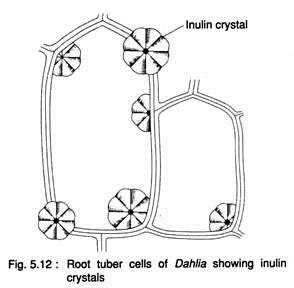
It is a polysaccharide that forms a powder-like compound and occurs in colloidal condition in the cell-sap of the vacuoles of plants like Dahlia, Artichoke etc. of the family Compositae. It is precipitated by treatment with alcohol or glycerine.
When blocks of root tubers of Dahlia are allowed to remain in alcohol or glycerine for some time and the sections are examined under the microscope, beautiful fan-shaped crystals are found inside the cells (Fig. 5.12). On hydrolysis, inulin yields fructose only. Inulin solution turns yellowish-brown when treated with acid phloroglucin. On precipitation, the crystals are easily recognised.
(ii) Starch grains:
Starch grains are the common insoluble carbohydrates. They are the most important reserve foods of the plants and occur in almost all the green plants excepting some algae. Starch grains may occur in all parts of the plants, but they are more abundant in the storage organs, cereal endosperms, fruits and seeds. They are very complex chemically with the formula, (C6H10O5)n where the value of n is not clearly known.
The building blocks of the molecule are unknown number of hexose sugars and is, thus, a polysaccharide. It is the stored food and is hydrolysed enzymatically into component hexoses when required.
There are two types of starch grains: assimilatoty starch and reserve starch. Assimilatory starch grains are temporary bodies formed by the chloroplasts in daytime during photosynthesis. They are granular in appearance and temporary in duration, as they are reconverted into sugar with nightfall.
Surplus sugars are transported to the sink and there they are converted into permanent starch grains to be stored up for future use by the colourless leucoplasts—the amyloplasts. These are reserve starch. They occur profusely in storage organs like roots and underground stems and in seeds and fruits.
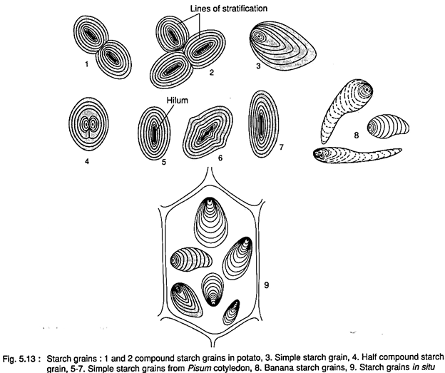
These starch grains are of diverse size and shape. Some of them are large enough to be seen even with the naked eye, whereas most of them are microscopic. Every starch grain has a shiny refractive point, known as hylum, which is the starting point.
Starch molecules are deposited in layers around the hilum giving rise to a distinct stratified appearance. The layers are referred to as lines of stratifications (Fig. 5.13). The starch grains are regarded as spherocrystals consisting of layers of sugar units arranged in a regular space-lattice.
Starch grains vary considerably in shape and size, depending on the source (Fig. 5.13). They are usually oval or elliptical in potato varying from 7 to 10nm in diameter, 3-4.5 nm in wheat, 1.2 to 1.8 nm in maize.
But the shape is more or less constant in a particular species. High amount of starch is accumulated mainly in storage organs. Thus potato tuber contains 15- 30% starch of its dry weight. The cereals contain 50 to 70% starch of their dry weight.
(iii) Glycogen:
It is another insoluble polysaccharide with the formula (C6H10O5)n. Though it is of abundant occurrence in animals, it also occurs in blue-green algae, slime moulds, fungi and bacteria as reserve foods. It is ultimately broken down to glucose by enzyme action and utilised as such. It gives reddish coloration with iodine solution.
Hemicellulose:
The reserve cellulose is called hemicellulose. Cellulose [(C6H10O5)n], is another polysaccharide which is much more complex than starch. The cell wall is primarily made up of cellulose. It is insoluble in water but readily soluble in alkalis, and is also indigestible.
In the seeds of many plants like date palm, ivory nut palm etc., another insoluble carbohydrate resembling cellulose is deposited as extra layers making the wall very thick and hard. This is reserve cellulose or hemicellulose.
(iv) Pectins:
Pectins are called fruit jellies and are present mainly in many fruits. They are readily soluble in water. Pectins can increase the water-holding capacity of the cells. The middle lamella is made up of pectic compound. They are abundantly used in the preparation of jams and jellies.
(v) Gums and Mucilages:
Gums and mucilages are widely found in the plants. They do not readily dissolve in water, but they form a slimy mass when moistened. Plants use them as reserve foods. Gums and mucilages also help the plants to hold water and prevent from desiccation. They facilitate dispersal of seeds. To human beings they are particularly useful in industries and medicine.
(b) Nitrogenous matters:
Proteins are the most common nitrogenous matters found in plants. They are the most complex organic macro- molecules made up of amino acids. They are required in large amounts in the regions of active growth. They serve as an essential component of protoplasm. Chemically, the proteins are composed of carbon, hydrogen, oxygen and nitrogen, and often sulphur, and, sometimes phosphorus.
Chlorophyll, the most essential green pigment of plants, is a magnesium-containing lipid and in association of protein it becomes functional. The building blocks of the protein molecules are the amino acids. If the amino acids are further aminated they form amides. Very common amides are asparagine, glutamine etc.
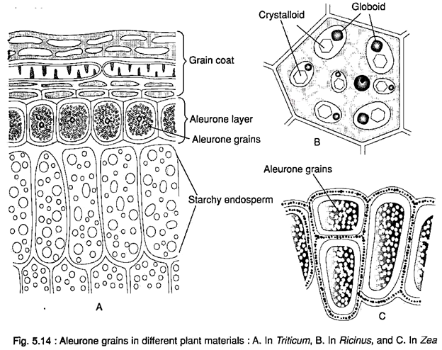
The stored proteins inside the cell are called proteid grains. Very common such proteid grains are gliadin found in wheat, zein in corn etc. In the storage regions they occur as grains called aleurone grains. Many plant proteins have been found to occur in crystalline or amorphous forms.
In cereals, a layer of cells just beneath the seed coat, composed of pericarp and testa, remains filled with aleurone grains and called aleurone layer. In pulses they remain as small grains mixed with comparatively latter starch grains.
More complex aleurone grains are found in the endosperm of castor seed in which the grain has a protein matrix. In that matrix there occurs a large crystalline Body, “called crystalloid, and a small round body, called globoid. The crystalloid is nitrogenous and the globoid is double phosphate of calcium and magnesium.
(c) Fats and Oils (= Triglycerides or triglycerols):
Fats and oils are also very important energy-storage reserve materials of plants along with carbohydrates and proteins. They are composed of carbon, hydrogen and oxygen having much more of carbon and hydrogen and relatively very low oxygen.
Chemically, triglycerides are esters of glycerol with three fatty acid molecules. Due to occurrence of relatively low oxygen and high carbon and hydrogen — fats and oils release much more energy than carbohydrates. Fats which are liquids at room temperature are called oils.
They are lighter than water, their specific gravity ranges from 0.875 to 0.970, and are insoluble in water. The physical characteristics of a triglyceride are determined by the chain length of the constituent fatty acids — whether the fatty acids are saturated or unsaturated.
The fats contain saturated fatty acids whereas the oils contain unsaturated fatty acids. Fats and oils dissolve in solvents like chloroform, ether, acetone, benzene etc. Oils are present in all living cells, often in form of droplets in the cytoplasm.
Triglycerides are esters of fatty acids and glycerol. Principal fatty acids are oleic acid, palmitic acid, capric acid, stearic acid, linoleic acid etc. They are abundantly present in storage organs, in seeds, embryos and meristematic tissues. They are formed in the cytosol and also by special fat-forming plastids called elaioplasts.
Fats usually occur in the plants as oils. These oils are non-volatile and, hence, are called fixed oils. Some oils are also used as medicines, like chaulmoogra oil used in leprosy treatment and croton oil used as one of the most strong purgatives.
Besides their utility as source of food, fats are also used in many industries. If fatty acids are saponified when treated with caustic soda or caustic potash — soap is formed.
(ii) Secretory Materials:
The secretory materials are not directly concerned with the nutrition and growth of the plants. They are often present in special sacs or glands.
The secretory materials are of the following types:
a. Enzymes:
Enzymes are the biocatalysts secreted by the protoplasm of many cells. These are proteinaceous, colloidal substances. They can catalyse the break-down of complex food materials into their simpler, soluble and diffusible forms. Enzyme action is usually specific and reversible.
Large number of enzymes is present in the plants. Apart from their role in digestion they regulate or control all biochemical processes inside the plants.
b. Nectar:
It is a sweet substance usually secreted by the floral parts of many plants. It is chiefly composed of sucrose, together with glucose and fructose, and is an excellent nutritious food. It is also used as medicines.
Nectar attracts a large number of insect visitors of pollination. The insects search for nectar in different flowers and carry pollen grains from one flower to the stigmas of other flower, thus they bring about pollination in many plants.
(iii) Excretory materials:
These ergastic substances are hardly of any use to the plants, so these metabolic by-products are called excretory materials or waste products. Most of these substances are very useful to mankind. As the rate of metabolism in plants is less the waste products formation is also lesser in quantity.
Moreover, the plants, unlike animals, have no definite excretory systems, excretory matters accumulate somewhere inside the plant- body. The plants, however, eliminate the waste products to some extent through leaf-falling and through fruits, feeds and barks of trees. Otherwise the accumulation of waste products would create toxicity and interfere with the activities of the living cells.
The common excretory materials are:
a. Alkaloid:
The term alkaloid, coined in 1819 in Halle, Germany, by a pharmacist, Carl Meissner, finds its origin in the Arabic name al-qali, the plant from which soda was first isolated. Alkaloids were originally defined as pharmacologically active, nitrogen-containing basic compounds of plant origin. Thus alkaloids are nitrogenous waste products found in many plants.
Chemically, they are composed of carbon, hydrogen, oxygen and nitrogen. They occur in storage organs like roots and seeds, in bark, leaves, wood and other parts. Alkaloids are usually insoluble in water, but readily soluble in alcohol. They have bitter taste, and many of them are deadly poisonous.
Alkaloids are the active principles of a large number of medicines derived from the plants. Many of them are still in use today as prescription drugs. One of the best- known prescription alkaloids is the antitussive and analgesic codeine from the opium poppy.
Plant alkaloids have also served as models for modern synthetic drugs, such as the tropane alkaloid atropine or tropicamide used to dilate the pupil during eye examination and the indole-derived antimalarial alkaloid quinine or chloroquine.
Quinine present in Cinchona bark, reserpine in the root of Rauwolfia serpentina, strychnine in nux vomica seeds, cocaine in coco, caffeine in coffea seeds, ephedrine in Ephedra, nicotine in tobacco, are the common alkaloids.
In addition to having a major impact on modern medicine, alkaloids have also influenced world geopolitics. Notorious example is the Opium Wars between Britain and China (1839 – 1859) and the efforts currently underway in various countries to eradicate illicit production of heroin, a semisynthetic compound derived by acetylation of morphine, and cocaine, a naturally occurring alkaloid of coca plant.
The physiologically active alkaloids participate in plant chemical defenses. It is supported by their wide range of physiological effects on animals and by the antibiotic activities many alkaloids possess. Various alkaloids also are toxic to insects or function as feeding deterrents.
b. Tannins:
‘Tannins’ is a collective term for a variety of plant polyphenols used in the tanning of raw hides to produce leather. They are widely distributed in plants and occur in especially high amounts in the bark of certain trees (e.g., oak) and in galls. These are usually bitter and astringent substances.
They occur in the cell-sap of the vacuoles, in the cell wall, in bark and woody parts of plants. Tannins are abundantly present in many fruits like myrobalans, in leaves, seeds, bark and also in pathological galls. The percentage of tannin is usually high in unripe fruits.
Tannins are classified as condensed and hydrolysable tannins. The condensed tannins are flavonoid polymers and are thus products of phenylpropanoid metabolism. The hydrolysable tannins consist of glycosylated gallic acids. Many of these gallic acids are linked to hexose molecules. Gallic acid in plants is formed mainly from shikimate.
The phenolic groups of the tannins bind so tightly to proteins — by forming hydrogen bonds with the -NH groups of peptides — that these bonds cannot be cleaved by digestive enzymes.
In the tanning process, tannin binds to the collagen of animal hides and thus produces leather which is able to withstand the attack of degrading microorganisms. Tannins turn blue black in colour in contact with iron salts. Thus they are extensively used for manufacture of ink. Some of them have medicinal value as well.
Tannins have unpleasant taste. They bind with the enzymes of saliva and thus the animals are discouraged from eating plants containing tannin. Tannins also react with enzymes of the herbivore digestive tract. For these reasons tannins are very effective in protecting leaves from being eaten by animals. So it contributes to the defense mechanism of the plants.
c. Latex:
Latex is usually a milky or often watery fluid present in some families of angiosperms. It is an emulsion of various substances like proteins, gums, resins, alkaloids, etc. remain suspended in an aqueous background. Dumbbell-shaped starch grains are frequently noticed in latex. It is useful to the plants in healing of wounds. This milky fluid is very valuable as the chief commercial source of rubber.
The majority of plants yielding rubber belong to the families Moraceae, Euphorbiaceae and Apocynaceae. Hevea brazilensis of family Euphorbiaceae needs special mention as a source of rubber.
Latex of papaya contains papain, the enzyme helpful in digestion of protein food. The so-called ‘Cow- tree’ Brosimum galactodendron of family Moraceae of Venezuala yields the latex which is taken by the natives as a substitute of milk.
d. Mineral Crystals:
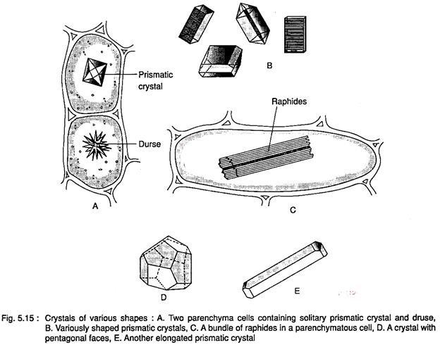
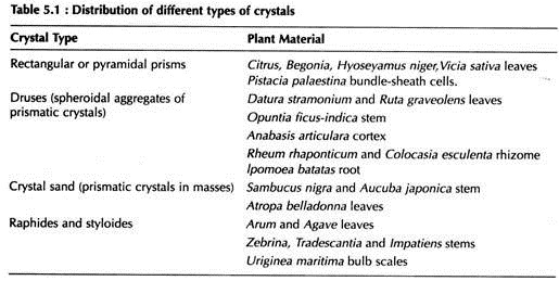
Many plants deposit in their cells crystals of various chemical nature consisting mostly of salts of calcium, chiefly calcium oxalate, and silicon dioxide. Crystals of many organic substances, such as berberin, saponin etc., are frequent. Any part of the plant body may contain crystals, still they are more abundant in the pith, cortex, and phloem. There are many structural forms of crystals found in plant cells (Fig. 5.15).
They may be solitary, rhombohedral, prismatic, pyramidal etc. Sheaflike bundles of long acicular crystals known as raphides, and clustered crystals in spheroidal masses called druses are most common. The individual crystals of druses may be raphides. Solitary raphides, small prismatic crystals, and minute crystals called crystal sand.
Usually one form of crystal may occur in a given cell. Two or more types may be found in the some cases. Many inorganic crystals appear to involve organic matter in the course of their formation. The larger crystals of xylem often show this condition, and druses frequently contain a prominent organic center.
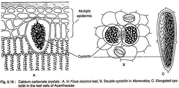
Calcium carbonate crystals usually occur in the epidermis impregnating a portion of cellulose cell wall in many plants. In rubber or banyan these Ca-carbonate crystals aggregate on a peg like projection of the cell wall looking like a bunch of grapes. The epidermis here is multi- seriate and the crystal-aggregation takes place in some enlarged cells of the innermost layer. Such a crystal-aggregate is called cystolith (Fig. 5.16).
e. Essential oils:
These are volatile oils found in many parts of the plant. They usually occur in special glands. The fragrant essence of many flowers and leaves, rinds of some fruits, bark and wood of some trees and even of some seeds are due to presence of essential oils.
These oils are advantageous to the plants in bringing about pollination and dispersal of fruits and seeds by attracting insect visitors. They are used in manufacture of perfumes, soaps, etc., and also in medicine, e.g., eucalyptus oil from Eucalyptus globulus of family Myrtaceae and valerian from Valeriana officinalis of family Valeianaceae. Essential oils also have antiseptic value.
f. Resins:
A resin is a polymer with a rigid three-dimensional network of phenol-formaldehyde repeating units. They occur in special glands or ducts usually in combination with gums and essential oils. They are water insoluble but soluble in many solvents like ether and alcohol. Resins are of three types. Pure resins which are secreted atone are usually solid and brittle, called hard resins.
They readily dissolve in alcohol and are used in paint and varnish industry as waterproofing and glossy agent. Lacquer and shellac are the examples. Resins mixed with essential oils are aromatic and more or less liquid in nature, known as oleo-resins, e.g., turpentines and Canadabalsam. The third type is gum resins which are mixtures of resins and gums. They are usually milky in appearance and collected by making an injury on the plant body. Many resins are used in medicine.
g. Gums:
Gums are metamorphosed, unorganized, amorphous products of organized cell- wall materials. Gums may also be formed from starch. The process of gum formation is called gummosis. During this process disintegration of the walls starts in the primary wall and proceeds towards the innermost lamella of the secondary cell-wall. It also occurs in the bark. Gums are water soluble.
They are exuded from the plants in natural course or through the injuries as mucilaginous masses. They are of great commercial value as adhesives, and are also used in other industries. Gumarabic of Acacia Senegal and other Acacia species is formed in the bark. It is used in medicine as an emulsifier.
