Are you looking for an essay on ‘Golgi Apparatus’? Find paragraphs, long and short essays on ‘Golgi Apparatus’ especially written for school and college students.
Essay on Golgi Apparatus
Essay Contents:
- Essay on the Introduction to Golgi Apparatus
- Essay on the Structure of Golgi Apparatus
- Essay on the Morphology and Chemistry of Golgi Apparatus
- Essay on the Functions of Golgi Apparatus
Essay # 1. Introduction to Golgi Apparatus:
In 1897, Camillo Golgi, who was investigating the nervous system by using a new self- developed staining technique, observed a cellular structure that he termed the internal reticular apparatus under his light microscope, which is later universally known as the Golgi apparatus. The Golgi apparatus found universally in both plant and animal cells, is typically comprised of a series of five to eight cup-shaped, membrane-covered sacs called cisternae that look something like a stack of deflated balloons.
The Golgi apparatus is often considered the distribution and shipping department for the cell’s or more or less the “postal office” of the cell. It process all incoming lipids, proteins, etc., and controls their export as well. These strangely shaped organelles act as a molecular sorting station, directing many of the proteins and carbohydrates used by the body to their correct locations after tagging them with structural modifications and destination information.
There are views that Golgi complex has originated from the plasma membrane or from nuclear envelope or from the annulated lamella. However, most of the workers believe in its origin from the ER, particularly from the rough ER by the loss of ribosome.
Essay # 2. Morphology and Chemistry of Golgi Apparatus:
The Golgi apparatus is morphologically very similar in both plants and animal cells but the number of Golgi complex in a cell is variable. The size of the Golgi varies with the metabolic state of cell and hence it is called as pleomorphic. It hypertrophies during hyper-function but reduces considerably during hypo-function. The Golgi complex generally lies between the nucleus and periphery usually showing specific orientation.
The Golgi complex is a lipoproteinaceous structure. It also has very low levels of nucleic acid and polysaccharides. Morphologically, the Golgi stands between ER and plasma membrane. The main enzymes of Golgi are thiamine pyrophosphatase (TPPase) and glycosyl transferase, the latter being most characteristic. The Golgi membrane has a single electron transport chain containing cytochrome b5, the enzyme acyl transferase and choline phosphotransferase are absent in Golgi membrane thus differing from ER.
Essay # 3. Structure of Golgi Apparatus:
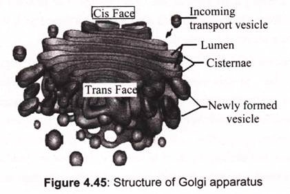
Dalton and Felix (1954) elucidated the structure of Golgi complex, which consist of three membranous components- A Golgi cisternae, Golgi vesicles and Golgi vacuoles. All the three structures are bound by a single unit membrane of 70Å thickness. The Golgi apparatus consists, like the ER of membranous structures. It is made up of a stack of flattened cisternae and similar vesicles. The Golgi cisternae or saccules or lamillar units are flattened membrane bound structures placed one over the other.
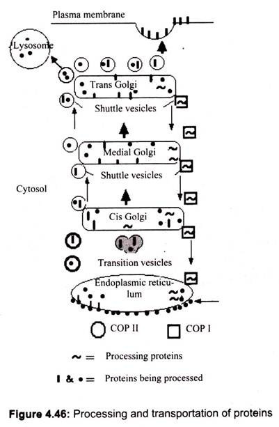
The number of cisternae in a complex generally varies from 3-8 but rarely there may be as many as 30 cisternae (Fig. 4.46). The distance between two unit membranes in a cisternae is about 150 Å. Its cis face is the side facing the ER, while the trans face is directed towards the plasma membrane. The cis and trans faces have different membranous compositions (Fig. 4.45).
In between Golgi cysternae, is found dense inter-cisternal material. The inter-cisternal material may be elongated measuring 60-80 Å in diameter or granular. Perhaps it is a cementing material which holds the cisternae together. Golgi vesicles pinched off from cisternae have a dimension of400-800 Å diameter.
A Golgi complex shows polarity having a proximal or forming face and a distal or maturing face. While the forming face shows convexity, the maturing face exhibits concavity. The forming face is generally closer to nuclear envelope or ER. The forming face is characterized by presence of certain transition vesicles or tubules converging upon the Golgi cisternae. Thus, in a surface view the Golgi cisternae appear reticulate or fenestrated. These transition vesicles are thought to give rise to the ER associated with the maturing face is a region consisting of smooth ER and developing lysosome.
Essay # 4. Functions of Golgi Apparatus:
Golgi apparatus is responsible for handling the macromolecules that are required for proper cell functioning. It processes and packages these macromolecules for use within the cell or for secretion. Primarily, the Golgi apparatus modifies proteins that it receives from the rough endoplasmic reticulum; however, it also transports lipids to vital parts of the cell and creates lysosomes. Some of the modifications made inside the Golgi complex include: attaching polysaccharides to proteins to form glycol-proteins, cutting proteins into smaller active fragments, incorporating phosphates onto protein molecules and addition of a sulfate group to molecules.
Other functions of the Golgi apparatus include the production of glycosaminoglycans, which go on to form parts of connective tissues. The Golgi will use a xylose link to polymerize the glycosaminoglycans onto proteins to form proteoglycan. It then performs sulfation onto the proteoglycans in order to aid in signaling abilities and giving the molecule a negative charge. The Bcl-2 genes that are located within the Golgi apparatus also play a significant role in preventing apoptosis, or the destruction of the cell. The Golgi complex in plant cells produces pectins and other polysaccharides specifically needed by for plant structure and metabolism.
In the endoplasmic reticulum, proteins synthesized by ribosomes are sent through the canals of the ER, where they meet up with the Golgi bodies. The proteins are then packaged in vesicles. The membranes of these vesicles are then able to bond with the cell membrane, where their contents are secreted outside the cell.
The transport vesicles from the ER fuse with the cis face of the Golgi apparatus (to the cisternae) and empty their protein content into the Golgi lumen (the internal space of the Golgi apparatus). The proteins are then transported through the Golgi apparatus towards the trans face and are modified on their way. The transport mechanism itself is not yet clear; it could happen by cisternae progression (the movement of the apparatus itself, building new cisternae at the cis face and destroying them at the trans face) or by budding (small vesicles transport the proteins from one cisterna to the next, while the cisternae remain unchanged).
Once the proteins reach the trans face, they are embedded into transport vesicles and brought to their final destinations. For example in the formation of glycol-proteins used in cell membranes proteins in vesicles from the ER will be directed to Golgi apparatus. In the Golgi apparatus carbohydrates are attached to them creating glycol-proteins. After they have been secreted in to the cell, the vesicles fuse to the cell membrane and release their contents (Fig. 4.46).
The cisternal maturation model states that the vesicles fuse to each other at the cis face of the Golgi apparatus and are essentially pushed along as new vesicles fuse together behind them. Newly synthesized proteins traverse the Golgi stack until they reach the trans-most Golgi compartment, which is termed the trans-Golgi network. The trans-Golgi network sorts the proteins into several types of vesicles. Using a variety of signals, the Golgi separates the products from the processing enzymes that made them and returns the enzymes back to the endoplasmic reticulum. Clathrin-coated vesicles carry certain proteins to lysosomes. Other proteins are packaged into secretory vesicles for immediate delivery to the cell surface. Still other proteins are packaged into secretory granules, which undergo regulated secretion in response to specific signals. This sorting function of the Golgi apparatus allows the various organelles to grow while maintaining their distinct identities.

