
Are you looking for an essay on ‘Meiosis’? Find paragraphs, long and short essays on ‘Meiosis’ especially written for school and college students.
Essay on Meiosis
Essay Contents:
- Essay on the Introduction to Meiosis
- Essay on the Process of Meiosis
- Essay on the Divisions of Meiosis
- Essay on the Significance of Meiosis
1. Essay on the Introduction to Meiosis:
Meiosis is a type of cell division that occurs only in eukaryotes, produces haploid (n) sex cells or gametes (which contain a single copy of each chromosome) from diploid (2n) cells (which contain two copies of each chromosome). In this process one DNA replication followed by two successive nuclear and cellular divisions (Meiosis I and Meiosis II). As in mitosis, meiosis is preceded by a process of DNA replication that converts each chromosome into two sister chromatids (Fig. 9.8).
The resulting four gametes are haploid, meaning that they contain half the number of chromosomes. This is the reason as to why meiosis cell division is also referred to as reduction division. During fertilization, two gametes, one from the mother and another from the father, fuse, thus resulting in doubling of chromosome number. The fusion of gametes leads to the production of a zygote that has the same chromosome number of the parents. Variation occurs in the resulting zygote due to the process of meiosis and fertilization of gametes. Zygote after attaining maturity is capable of dividing into daughter cells.

There are two different sex cells or gametes- sperm and eggs. Males produce sperm which females produce eggs because they are produced from germ cells, gametes are likewise haploid. In order to create a new individual via sexual reproduction, a sperm cell needs to activate an egg by joining it in a fertilization process. These two haploid cells can unite in to a diploid cell, which can then develop into a new individual. The sexual reproductive processes ensure the resulting offspring will have an equal maternal and paternal genetic contribution.
Life cycles of all sexually-reproducing organisms follow this pattern of alternation of generations. The 2n adult produces 1n gametes by the process of meiosis. These unite in the process of syngamy to produce a new 1n generation. Thus, the life cycles alternate between 1n and 2n stages, and between the processes of meiosis and syngamy. Meiosis was discovered and described for the first time in sea urchin eggs in 1876, by a German biologist Oscar Hertwig (1849-1922). It was described again in 1883, at /he level of chromosomes, by Belgian zoologist Edouard Van Beneden (1846-1910), in Ascaris worms’ eggs. In animals meiosis helps in the production of the gametes: sperni and eggs, while in plants used to produce spores.
The process of meiosis is exhibited by higher forms of organisms, which reproduce sexually. In plants, meiosis is observed after spore .production; whereas, in animals, meiosis takes place during gamete (sperm and egg) formation. Like other cell division, meiosis produces daughter cells. However, there is a significant difference between meiosis and other types of cell division like mitosis or binary fission. In meiosis, the parent cell divides and produces four gametes that are not capable of further division; whereas, in other types of cell division, the parent cell produces identical daughter cells, which can undergo division on their own.
2. Essay on the Process of Meiosis:
The steps in meiosis are similar to mitosis and even known by having the same names. However, there is a significant difference in regions to the way the chromosomes line up initially. In mitosis, chromosomes line up individually, while in meiosis, the two chromosomes in each homologous pair line up next to each other as pairs. This pairing process is called synapsis, and the resulting homologous pair is called a bivalent in reference to the two chromosomes or a tetrad in reference to the four sister chromatids involved.
There are two stages of meiosis, namely, meiosis I and meiosis II. The parent cell or the dividing cell undergoes a preparatory phase, known as interphase, before entering the two stages of meiosis. In the interphase, the parent cell synthesizes more DNA (deoxyribonucleic acid) and proteins, increasing the overall size and mass of the cell. As a part of the preparatory phase, the dividing cell duplicates or doubles its chromosomes. With these major changes, the parent cell enters the first stage of meiosis.
Prior to meiosis, all chromosomes are duplicated in a process similar to chromosomes duplication prior to mitosis. Outside the nucleus of animal cells there are two centrosomes, each containing a pair of centrioles. The two centrosomes are produced by the duplication of a single centrosome during premeiotic interphase. The centrosomes serve as microtubule organizing centers (MTOCs). Microtubules extend radially from centrosomes, forming an aster. PI ant cells do not have centrosomes.
3. Essay on the Divisions of Meiosis:
i. First Division of Meiosis (Meiosis I):
(a) Prophase I:
In beginning of prophase I, the chromosomes have already duplicated, and they coil and become shorter and thicker and visible under the light microscope (Fig. 9.9). The duplicated homologous chromosomes pair, and crossing-over (the physical exchange of chromosome parts) occurs. Crossing-over is the process that can give rise to genetic recombination. At this point, each homologous chromosome pair is visible as a bivalent (tetrad), a tight grouping of two chromosomes, each consisting of two sister chromatids. The sites of crossing-over are seen as crisscrossed non-sister chromatids and are called chiasmata (singular: chiasma). The homologous chromosomes pair and exchange DNA, to form recombinant chromosomes.

Prophase I is divided into five phases:
(i) Leptotene:
Chromosomes start to condense. Homologous dyads (pairs of sister chromatids) find each other and align themselves from end to end with the aid of an axial element (that contains cohesins). In budding yeast (and perhaps other eukaryotes) the process follows a period of trial-and-error. Any two dyads pair at their centromeres. If they are not homologs, they separate and try again. Double-stranded breaks (DSBs) often occur in the DNA of the chromatids, and these may be necessary for the homologs to recognize each other.
(ii) Zygotene:
Homologous chromosomes become closely associated (synapsis) to form pairs of chromosomes (bivalents) consisting of four chromatids (tetrads). The pairing of homologous chromosomes is known as synapsis (Gr. Synapsis= union). The synapsis begins at one or more points along the length of the homologues chromosomes. Three types of synapsis have been observed preterminal, precentric and localized on the basis of position of synapsis. The paired homologous chromosomes are joined by a roughly 0.2 µm thick protein containing synaptonemal complex (SC). Synaptonemal complex helps stabilize the pairing of homologous chromosomes and to facilitate the cytogenetical activity called crossing over or recombination.
(iii) Pachytene:
Crossing over takes place between pairs of homologous chromosomes to form chiasmata (sing. chiasma). They are named for the idea that they represent points where DNA recombination is occurring. There must be at least one for each bivalent if meiosis is to succeed. There is often more, each one presumably representing the point of a crossover. They contain enzymes known to be needed for DNA recombination and repair. The steps in recombining DNA continue to the end of pachytene.
(iv) Diplotene:
Homologous chromosomes start to separate but remain attached by chiasmata. DNA recombination is complete. The synaptonemal complex begins to break down. The chromatids begin to pull apart revealing chiasmata. At first the chiasmata are located at the sites of the recombination nodules, but later they migrate towards the ends of the chromatids.
(v) Diakinesis:
Homologous chromosomes continue to separate, and chiasmata move to the ends of the chromosomes. In some organisms, the chromosomes decondense and begin to be transcribed for a time. This is followed by the chromosomes recondensing in preparation for metaphase I. In creatures where this does not occur, the chromosomes condense further in preparation for metaphase I.
(b) Metaphase I:
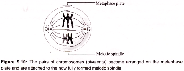
The centrioles are at opposite poles of the cell. The pairs of homologous chromosomes (the bivalents), now as tightly coiled and condensed as they will be in meiosis, become arranged on a plane equidistant from the poles called the metaphase plate. Spindle fibers from one pole of the cell attach to one chromosome of each pair (seen as sister-chromatids), and spindle fibers from the opposite pole attach to the homologous chromosome (again, seen as sister chromatids).
(c) Anaphase I:
Anaphase I begin when the two chromosomes of each bivalent (tetrad) separate and start moving toward opposite poles of the cell as a result of the action of the spindle. In anaphase I the sister chromatids remain attached at their centromeres and move together toward the poles. A key difference between mitosis and meiosis is that sister chromatids remain joined after metaphase in meiosis I, whereas in mitosis they separate (Fig. 9.11).

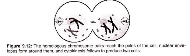
(d) Telophase I:
In Telophase I the homologous chromosome pairs complete their migration to the two poles as a result of the action of the spindle. Now a haploid set of chromosomes is at each pole, with each chromosome still having two chromatids. A nuclear envelope reforms around each chromosome set, the spindle disappears, and cytokinesis follows. In animal cells, cytokinesis involves the formation of a cleavage furrow, resulting in the pinching of the cell into two cells (Fig. 9.12). After cytokinesis, each of the two progeny cells has a nucleus with a haploid set of replicated chromosomes.
ii. Second Division of Meiosis (Meiosis II):
Gamete Formation:
(a) Prophase II:
Meiosis II begins without any further replication of the chromosomes. In prophase II, the nuclear envelope breaks down and the spindle apparatus forms. Here, the centrioles duplicate. This occurs by separation of the two members of the pair, and then the formation of a daughter centriole perpendicular to each original centriole. The two pairs of centrioles separate into two centrosomes. The nuclear envelope breaks down, and the spindle apparatus forms.
(b) Metaphase II:
The chromosomes become arranged on the metaphase plate, much as the chromosomes do in mitosis, and are attached to the now fully formed spindle (Fig. 9.13).

Each of the daughter cells completes the formation of a spindle apparatus. Single chromosomes align on the metaphase plate, much as chromosomes do in mitosis. This is in contrast to metaphase I, in which homologous pairs of chromosomes align on the metaphase plate. For each chromosome, the kinetochores of the sister chromatids face the opposite poles, and each is attached to a kinetochore microtubule coming from that pole.
(c) Anaphase II:
The centromeres separate and the two chromatids of each chromosome move to opposite poles on the spindle. The separated chromatids are now called chromosomes.
(d) Telophase II:
In Telophase II a nuclear envelope forms around each set of chromosomes. Cytokinesis takes place, producing four daughter cells (gametes, in animals), each with a haploid set of chromosomes because of crossing-over, some chromosomes are seen to have recombined segments of the original parental chromosomes.

4. Essay on the Significance of Meiosis:
Meiosis facilitates stable sexual reproduction. Without the halving of ploidy, or chromosome count, fertilization would result in zygotes that have twice the number of chromosomes as the zygotes from the previous generation. Successive generations would have an exponential increase in chromosome count. In organisms that are normally diploid, polyploidy, the state of having three or more sets of chromosomes, results in extreme developmental abnormalities or lethality. Polyploidy is poorly tolerated in most animal species.
Plants, however, regularly produce fertile, viable polyploids. Polyploidy has been implicated as an important mechanism in plant speciation. Most importantly, recombination and independent assortment of homologous chromosomes allow for a greater diversity of genotypes in the population. This produces genetic variation in gametes that promote genetic and phenotypic variation in a population of offspring.
The normal separation of chromosomes in meiosis I or sister chromatids in meiosis II is termed disjunction. When the separation is not normal, it is called nondisjunction. This results in the production of gametes which have either too or too a few of a particular chromosome, and is a common mechanism for trisomy or monosomy. Nondisjunction can occur in the meiosis I or meiosis II, phases of cellular reproduction, or during mitosis.
This is a cause of several medical conditions in humans (such as):
i. Down Syndrome – trisomy of chromosome 21
ii. Patau Syndrome – trisomy of chromosome 13
iii. Edward Syndrome – trisomy of chromosome 18
iv. Klinefelter Syndrome – extra X chromosomes in males – i.e. XXY, XXXY, XXXXY
v. Turner Syndrome – lacking of one X chromosome in females – i.e., XO
vi. Triple X syndrome – and extra X chromosome in females
Process of Gametogenesis:
Gametogenesis is the process of forming gametes (haploid, n) from diploid cells of the germ line. Spermatogenesis is the process of forming sperm cells by meiosis (in animals, by mitosis in plants) in specialized organs known as gonads (in males these are termed testes). After division the cells undergo differentiation to become sperm cells. Oogenesis is the process of forming an ovum (egg) by meiosis (in animals, by mitosis in the gametophyte in plants) in specialized gonads known as ovaries, whereas in spermatogenesis all 4 meiotic products develop into gametes, oogenesis places most of the cytoplasm into the large egg. The other cells, the polar bodies, do not develop. Human males produce 200,000,000 sperm per day, while the female produces one egg (usually) each menstrual cycle.
i. Spermatogenesis:
Sperm production begins at puberty and continues throughout life, with several hundred million sperm being produced each day. Once sperms form they move into the epididymis, where they are matured and stored (Fig. 9.15).

ii. Oogenesis:
The ovary contains many follicles composed of a developing egg surrounded by an outer layer of follicle cells. Each egg begins oogenesis as a primary oocyte. At birth each female carries a lifetime supply of developing oocytes, each of which is in Prophase I. A developing egg (secondary oocyte) is released each month from puberty until menopause, a total of 400- 500 eggs.